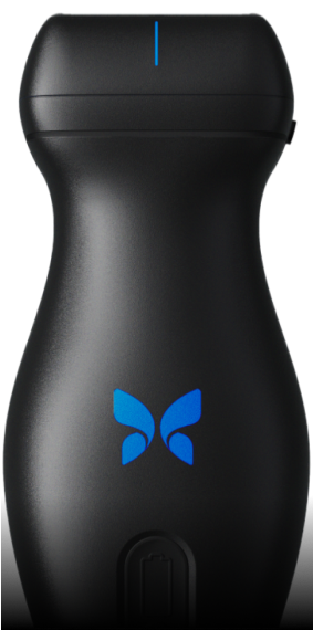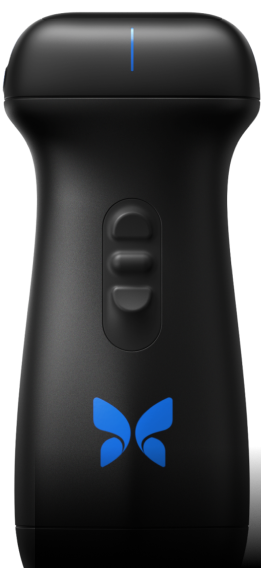
Hardware.
Compatibility.
Cleaning.
2. Disconnect the probe from the mobile device.
3. Wipe the probe, strain relief, cable, and connector with one of the recommended liquid saturated wipes for one (1) minute and until visibly clean.
4. Change the wipes as necessary and repeat the above step until the probe is visibly clean.
5. To dry the probe, use a soft cloth to blot the lens dry. Do not wipe the lens. Dry the rest of the probe, cable, strain relief, and connector.
6. Visually inspect the probe in a well-lit area to ensure all surfaces are clean. If the probe is not clean, repeat the cleaning steps above.
7. Dispose of cleaning material in accordance with all applicable regulations.
Imaging.
Presets.
This preset uses a curvilinear format to evaluate abdominal organs, such as the liver and kidney. It can also be used for abdominal procedures, such as abdominocentesis. It offers M-Mode and Color Doppler as well as linear and circumferential measurements.
Similar to our Abdomen preset, this preset was designed to provide better imaging in larger animals. It changes frequencies with depth to ensure consistent reliable visualization of up to 30cm.
This preset has been optimized for the contrast and detail required for bladder assessment. The Field of View (FOV) is expanded to 120 deg to enable visualization of the bladder in the near field. This includes the application of harmonic imaging, which uses resonant characteristics of tissue to produce imaging of higher resolution of the bladder wall, and surrounding pelvis. It provides Color Doppler carefully optimized for the identification of ureteral jets. It can also be used for collection procedures, such as cystocentesis.
This preset employs a high frame rate and applies a sector scan at higher frequencies to allow reliable real-time echocardiographic interrogation. M-Mode and Color Doppler are both supported.
This preset employs a high frame rate and applies a sector scan at higher frequencies to allow reliable real-time echocardiographic interrogation. Coherence Imaging, located in the Cardiac preset, improves contrast resolution by removing unmodeled acoustic reverberation typically known as “clutter.”
Designed for cardiac imaging in larger animals. Multiple parameters have been adjusted to provide for diagnostic quality echocardiography in patients needing more penetration. Coherence Imaging, located in the Cardiac Deep preset, improves contrast resolution by removing unmodeled acoustic reverberation typically known as “clutter.”
This preset employs a high frame rate and applies a sector scan at higher frequencies to allow reliable real-time echocardiographic interrogation. M-Mode and Color Doppler are both supported.
This preset has been designed with parameters best for imaging of the fast-moving dynamics of the heart in larger animals. It employs a high frame rate and applies a sector scan which allows reliable real-time echocardiographic interrogation. M-Mode and Color Doppler are both supported. Multiple parameters have been changed to adjust for this use case, such as using blended frequencies, to provide diagnostic quality echocardiography in patients needing more penetration.
Similar to Cardiac Deep, this preset was designed to provide more contrast with cardiac imaging for larger animals needing more penetration.
This preset is optimized to support imaging of lung pathology for animals in respiratory distress. In the very near field, a high-frequency linear format is optimized for the visualization of lung sliding. As the depth changes, so too does the image format and frequency range in order to support deeper interrogation for A or B Lines
This linear preset applies imaging parameters to display the fine balance of temporal, detail and contrast resolution required for high-frequency musculoskeletal exams and ultrasound-guided injections. This includes high-resolution imaging of muscles, tendons, ligaments, and bursae.
Similar to Musculoskeletal, this preset has been designed to increase the contrast resolution for a crisp visualization of the structures of interest via post-processing capabilities. Moreover, instead of a single focus and transmit frequency for the whole field of view, this preset divides the field of view into two parts at ≥ 5cm. A closer focus and higher frequency are applied in the near field.
Nerve imaging is a high frequency linear imaging format optimized for interrogation of peripheral nerves from a minimum depth of 1 cm to a maximum depth of 8 cm. It has been developed to show contrast between the nerve bundle distinct from surrounding structures, and yet maintain the the detail resolution required to demonstrate the typical “honeycomb” appearance of individual nerve fascicles
The Ophthalmic preset has been carefully developed with the appropriate MI (Mechanical Index) operating scope for point-of-care ultrasound examinations of the eye. This typically involves looking at structural integrity of the orbit and detection of foreign bodies, as well as retinal and optic nerve abnormalities.
This preset has been optimized to support the demonstration of soft tissue structures in the small parts anatomical category, such as bowel or pancreas. Focus has been made specifically to detail resolution and vascular sensitivity in a linear format.
This preset optimizes the image for top visualization of vascular structures, with a specific focus on peripheral arteries and veins in a linear format. It maximizes contrast resolution to provide a crisp demonstration of vascular borders and needle guidance.